Introduction
Magnetic Resonance Imaging (MRI) is a powerful diagnostic tool that has revolutionized the field of medical imaging. Unlike traditional X-rays or CT scans, MRI uses strong magnetic fields and radio waves to create detailed images of the inside of the body. This non-invasive technique allows doctors to see organs, tissues, and other structures with remarkable clarity, making it invaluable for diagnosing a wide range of conditions.
Brief Overview of MRI Technology
MRI technology works by aligning the protons in the body’s hydrogen atoms using a strong magnetic field. When these protons are exposed to radiofrequency pulses, they produce signals that are detected by the MRI machine. These signals are then converted into detailed images by a computer. The ability to adjust the magnetic field and radiofrequency pulses allows for different types of images, providing comprehensive views of various body parts.
Importance of Understanding MRI Scans
Understanding MRI scans is crucial for both patients and healthcare providers. For patients, knowing what to expect can alleviate anxiety and ensure a smoother experience. It also helps them make informed decisions about their healthcare. For healthcare providers, a deep understanding of MRI technology and its applications enables accurate diagnosis and effective treatment planning. Additionally, being aware of the potential side effects and safety considerations ensures that MRI scans are used appropriately and safely.
1. What is an MRI Scan?
Definition and Basic Principles
Magnetic Resonance Imaging (MRI) is a non-invasive imaging technique used to create detailed images of the organs and tissues within the body. Unlike X-rays and CT scans, which use ionizing radiation, MRI relies on powerful magnetic fields and radio waves to generate images. This makes MRI particularly useful for imaging soft tissues, such as the brain, muscles, and ligaments, which are not as clearly visible with other imaging methods.
How MRI Works: Magnetic Fields and Radio Waves
The process of MRI involves several key steps:
- Magnetic Field Alignment: The MRI machine generates a strong magnetic field that aligns the protons in the body’s hydrogen atoms. These protons are abundant in the human body, especially in water and fat.
- Radiofrequency Pulses: Once the protons are aligned, the machine sends out radiofrequency pulses. These pulses temporarily knock the protons out of alignment.
- Signal Detection: As the protons return to their original alignment, they emit signals. These signals are detected by the MRI machine’s receiver coils.
- Image Formation: The detected signals are processed by a computer to create detailed images of the body’s internal structures. By adjusting the magnetic field and radiofrequency pulses, different types of images can be produced, highlighting various tissues and abnormalities.
2. Common Uses of MRI Scans
Diagnosing Neurological Conditions
MRI scans are particularly valuable in the field of neurology. They provide detailed images of the brain and spinal cord, helping doctors diagnose conditions such as multiple sclerosis, stroke, brain tumors, and aneurysms. The high-resolution images allow for the detection of even small abnormalities, making MRI an essential tool for early diagnosis and treatment planning.
Imaging Soft Tissues: Muscles, Ligaments, and Organs
One of the key strengths of MRI is its ability to image soft tissues with exceptional clarity. This makes it ideal for examining muscles, ligaments, and organs. For instance, MRI can be used to diagnose torn ligaments, muscle injuries, and joint abnormalities. It is also commonly used to assess organs such as the liver, kidneys, and heart, providing detailed information about their structure and function.
Detecting Tumors and Abnormalities
MRI is highly effective in detecting tumors and other abnormalities within the body. It can differentiate between normal and abnormal tissues, making it easier to identify the presence of tumors, cysts, and other growths. MRI is often used in cancer diagnosis and staging, helping doctors determine the size, location, and extent of tumors. This information is crucial for developing effective treatment plans.
Monitoring Treatment Progress
MRI scans are also used to monitor the progress of treatment for various conditions. For example, in cancer patients, MRI can track the response to chemotherapy or radiation therapy, allowing doctors to adjust treatment plans as needed. Similarly, MRI can be used to monitor the healing process of injuries and the effectiveness of surgical interventions. This ongoing assessment helps ensure that patients receive the most appropriate and effective care.
3. The MRI Procedure: What to Expect

Preparation Steps Before the Scan
- Inform Your Physician: If you are claustrophobic, let your doctor know. They may prescribe a sedative to help you relax during the procedure.
- Metal Implants: Notify your doctor about any metal implants in your body, such as pacemakers, metal plates, or cochlear implants, as these can interfere with the MRI.
- Health Conditions: Discuss any health issues, such as kidney problems or allergies to contrast dyes, with your doctor.
- Medications: Continue taking your medications as usual unless instructed otherwise.
- Clothing and Accessories: Remove all metal items, including jewelry, watches, and hairpins. You may be asked to change into a hospital gown.
Step-by-Step Guide to the MRI Process
- Arrival: Upon arrival, you will fill out a patient history form and may be asked to change into a gown.
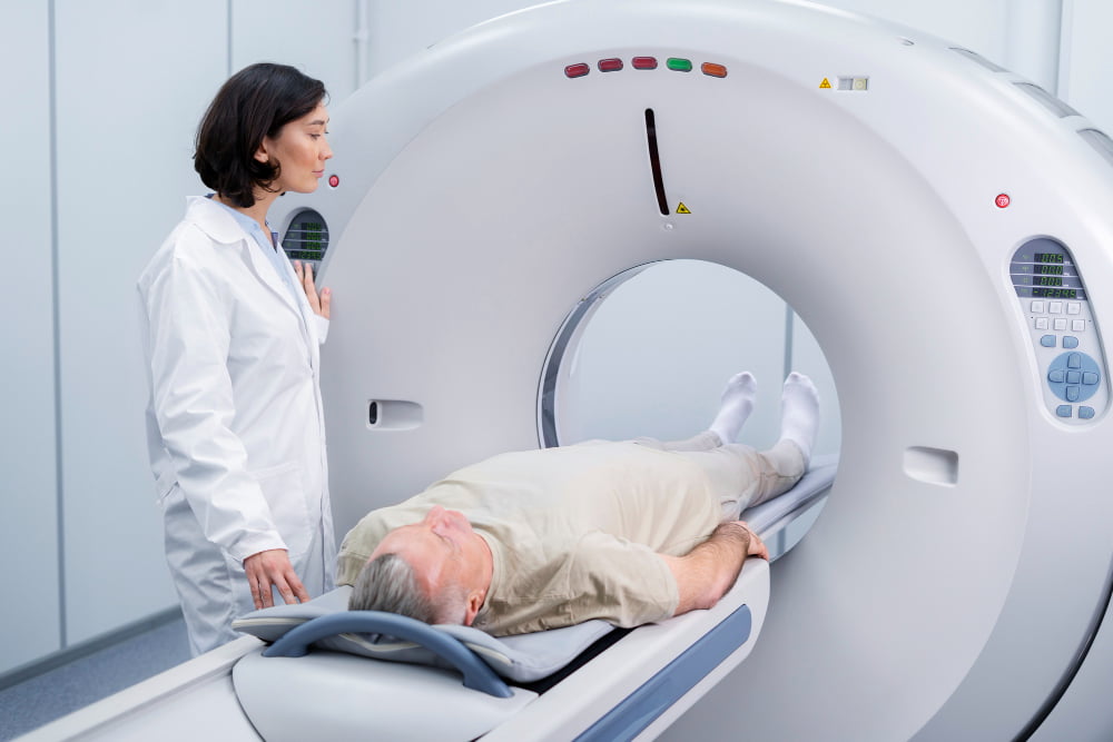
- Positioning: You will lie on a padded table that slides into the MRI machine, which looks like a large tube open at both ends.
- During the Scan:
- You must lie still to avoid blurring the images.
- You will be given earplugs to block out the loud thumping noises from the machine.
- A coil may be placed around the part of your body being scanned to improve image quality.
- The technologist will monitor you from another room and communicate with you through a microphone.
- Completion: The scan typically takes between 20 to 90 minutes, depending on the area being examined.
Duration and Comfort Tips
- Duration: The MRI scan can last from 15 to 90 minutes.
- Comfort Tips:
- Relaxation: Try to stay relaxed and breathe normally. Movement can distort the images.
- Communication: Use the squeeze button to alert the technologist if you feel uncomfortable at any time.
- Ear Protection: Wear the provided earplugs to reduce noise levels.
- Comfort Items: Some facilities allow you to bring a blanket or pillow for added comfort during the scan.
4. Benefits of MRI Scans
Non-invasive and Painless
MRI (Magnetic Resonance Imaging) scans are non-invasive, meaning they do not require any incisions or injections (unless a contrast dye is used). The procedure is painless, as it involves lying still in the MRI machine while it takes detailed images of the inside of your body. This makes it a comfortable option for patients who may be anxious about more invasive diagnostic procedures.
No Radiation Exposure
Unlike X-rays and CT scans, MRI scans do not use ionizing radiation. Instead, they use powerful magnets and radio waves to generate images. This makes MRI a safer option, especially for patients who need multiple scans over time, such as those with chronic conditions or those undergoing cancer treatment. The absence of radiation reduces the risk of potential long-term side effects.
High-Resolution Images for Accurate Diagnosis

MRI scans provide high-resolution images that offer detailed views of soft tissues, organs, and other internal structures. This level of detail helps doctors accurately diagnose a wide range of conditions, from torn ligaments and brain tumors to spinal cord injuries and heart disease. The clarity of MRI images can lead to more precise treatment plans and better patient outcomes.
MRI scans are a valuable tool in modern medicine, offering a safe, painless, and highly effective way to diagnose and monitor various health conditions. If you have any concerns or questions about undergoing an MRI, be sure to discuss them with your healthcare provider.
5. Potential Side Effects and Risks
Common Side Effects: Claustrophobia, Noise Discomfort
- Claustrophobia: Some patients may feel anxious or claustrophobic inside the MRI machine due to its enclosed nature. If you have a history of claustrophobia, inform your doctor beforehand. They may provide a sedative to help you relax during the scan.
- Noise Discomfort: MRI machines produce loud thumping and banging noises during the scan. Earplugs or noise-canceling headphones are usually provided to help reduce the discomfort. You can also listen to music if the facility offers this option.
Rare Side Effects: Allergic Reactions to Contrast Dye
- Allergic Reactions: In some cases, a contrast dye (usually gadolinium-based) is used to enhance the images. Although rare, some patients may experience allergic reactions to the dye. Symptoms can include itching, rash, nausea, or more severe reactions like difficulty breathing. Always inform your healthcare provider about any known allergies or previous reactions to contrast dyes.
Safety Considerations for Patients with Implants
- Metal Implants: Patients with metal implants, such as pacemakers, cochlear implants, or metal plates, need to inform their doctor before the MRI. The strong magnetic field can interfere with these devices, potentially causing harm or affecting the quality of the images.
- Other Implants: Some implants, like certain types of stents or artificial joints, maybe MRI-safe. However, it’s crucial to provide detailed information about any implants to your healthcare provider to ensure safety during the scan.
6. Tips for a Smooth MRI Experience
How to Prepare Mentally and Physically
- Mental Preparation:
- Stay Informed: Understanding the procedure can help reduce anxiety. Ask your healthcare provider any questions you have about the MRI process.
- Relaxation Techniques: Practice deep breathing, meditation, or visualization techniques to stay calm before and during the scan.
- Support: If you’re feeling particularly anxious, consider bringing a friend or family member for support.
- Physical Preparation:
- Diet and Hydration: Follow any specific instructions regarding eating or drinking before the scan. Generally, staying hydrated is beneficial.
- Clothing: Wear comfortable, loose-fitting clothes without metal fasteners. You may be asked to change into a hospital gown.
- Remove Metal Items: Take off all jewelry, watches, and other metal accessories before the scan.
Communicating with Your Healthcare Provider
- Discuss Concerns: Share any fears or concerns you have about the MRI with your healthcare provider. They can offer reassurance and solutions, such as sedatives for claustrophobia.
- Medical History: Provide a complete medical history, including any implants, allergies, or previous reactions to contrast dyes.
- During the Scan: Use the communication system in the MRI machine to speak with the technologist if you feel uncomfortable or need assistance.
Post-Scan Care and Follow-Up
- Immediate Care: After the scan, you can usually resume normal activities unless you were given a sedative. If so, arrange for someone to drive you home.
- Contrast Dye: If contrast dye is used, drink plenty of water to help flush it out of your system.
- Follow-Up: Schedule a follow-up appointment with your healthcare provider to discuss the results of the MRI. They will explain the findings and any next steps in your treatment plan.
By preparing mentally and physically, maintaining open communication with your healthcare provider, and following post-scan care instructions, you can ensure a smooth and stress-free MRI experience. If you have any specific questions or concerns, don’t hesitate to reach out to your healthcare team.
In conclusion, MRI scans are a powerful diagnostic tool that offers detailed images of the body’s internal structures without the risks associated with ionizing radiation. They are invaluable in diagnosing a wide range of conditions, from neurological disorders to musculoskeletal injuries. While generally safe, it’s important to be aware of potential side effects, such as allergic reactions to contrast dye and the rare discomfort some patients may experience. Understanding both the uses and side effects of MRI scans can help patients make informed decisions about their healthcare. Always consult with your healthcare provider to determine if an MRI is the right choice for your specific medical needs.


